Structural study of a protein in an ultrasound levitated drop
دراسة التركيب الفراغي لبروتين في قطرة معلقة بأمواج فوق-صوتية
The science of crystallography using x-ray diffraction to determine 3D-structure of simple and complex molecules is witnessing relatively new progress that rely on hanging droplets containing protein crystals (and any other molecules in crystalline state). The link below describes the structure determination of the Lysozyme by this revolutionary technique.
علم البلورات باستخدام إنكسار أو حيود الأشعة السينية x-ray diffraction لحساب وتحديد التراكيب الفراغية للجزيئات البسيطة والمعقدة منها يشهد تطورا جديدا نسبيا يعتمد على تعليق قطرات تحمل بلورات البروتينات (وأي نوع آخر من الجزيئات في حالة متبلرة). الرابط أدناه يصف كيف تم الحصول على التركيب ثلاثي الأبعاد لإنزيم الليزوزيم Lysozyme بهذه الطريقة المبتكرة.
This technique has many advantages over the usual methods as it allows for structural analysis in the room temperature, that is no need for ultra-freezing of the crystals as is the usual procedure, dispense with the crystal holder (necessary in the normal way) and saving towards the costs of the entire study.
لهذه التقنية جوانب إيحابية مهمة و منها إمكانية القيام بالتحاليل التركيبية في درجة حرارة الغرفة (لا حاجة للتبريد الفائف المستخدم عادة)، الإستغناء عن حامل البلورات الضروري في الطريقة العادية والإقتصاد في تكاليف الدراسة برمتها.
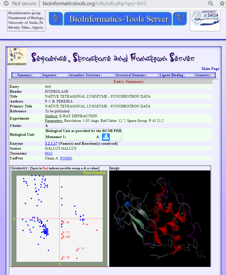
The determined structure done through this technique, see Image 3, can be obtained from the Protein Data Bank - PDB using the identification code 5FDJ. Exploration of the structure is available using the SSFS service, the link below:
التركيب الفراغي الذي تم الحصول عليه بهذه التقنية، الصورة المرفقة رقم 3، يتواجد في بنك/قاعدة البيانات المعروف بـ Protein Data Bank أو PDB يحمل الرمز 5FDJ و يمكن استكشافه علىى نظام الـ SSFS المتوفر على الرابط التالي:
(أنظر كذلك الصورة المرفقة رقم 4 .. . .. . .. . .. . .. . .. . .. See also Image 4)
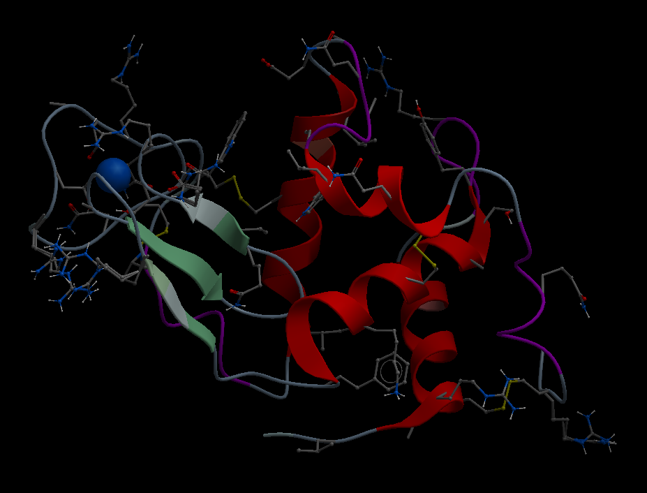
It remains, in our view, to carefully assess the effect of the ultrasound waves on the refined structures which seems missing in the article. The scientists behind the study confirm high conformational similarity of their obtained structure to the know structure of the lysozyme as shown in Images 5 and 6.
في رأينا الشخصي، يبقى أن يتم التأكد من مدى تأثير الأمواج الفوق-صوتية على التركيب الفراغي الناتج عن عمليات الحساب النهائية الأمر الذي يبدوا أنه لم يتم التركيز عليه في الموضوع المنشور وإن كان أصحاب البحث يؤكدون أن التركيب الهندسي لجزيئ الليزوزيم الذي تحصلوا عليه كان مطابقا بشكل كبير للتراكيب المعروفة سابقا لهذا الإنزيم (كما تبين الصورتين المرفقة رقم 5 و 6 لليزوزيم الذي تم حساب تركيبه سابقا بالتقنية التقليدية).
For more details, follow the article below:
للمزيد من التفاصيل تابع للموضوع الغير تقني على الرابط:
The technical paper published in the journal "Nature" is on the following link:
لقراءة البحث التقني في الموضوع والمنشور في المجلة المرموقة "Nature" على الرابط التالي:
See also the link below for more information:
أنظر كذلك الموضوع حول إنزيم الليزوزيم على الرابط:
🖝 Lysozyme
أجمل التحيات All the best


